Blood Vessels Labeled Head - Veins Arteries And Lymphatics Of The Face Dummies - Blood vessels labeled head :
byAdmin-
0
Blood Vessels Labeled Head - Veins Arteries And Lymphatics Of The Face Dummies - Blood vessels labeled head :. The largest arteries in the neck are the common carotids.these arise from the brachiocephalic trunk on the right side and directly from the arch of the aorta on the left. • arteries, which carry the blood away from the heart. In the neck and head exterior to the skull, the external carotid artery provides blood flow to the skin, muscles, and organs. The subclavian artery is divided into three parts based on anatomical landmarks. • capillaries, which enable the actual exchange of water and chemicals between the blood and the tissues.
Blood vessels labeled head : The common cartoid artery extends from the brachiocephalic artery. Blood vessels labeled head : Most head and neck blood drains through three vein pairs on each side of the neck that empty into the subclavian vein. The first part extends from its origin to the medial border of the scalenus anterior muscle.
What Are Blood Vessels With Pictures from images.infobloom.com The superior vena cava is the large vein that brings blood from the head and arms to the heart, and the inferior vena cava brings blood from the abdomen and legs into the heart. Blood vessels labeled head / major arteries of the head and neck carotid teachmeanatomy / these are the pulmonary and systemic circuits. Once blood is oxygenated in the lungs, it returns to the heart and is then pumped throughout the body. These include the common carotid artery that carries blood from the heart to the brain. Dimitrios mytilinaios md, phd last reviewed: Blood vessel lumen staining outperforms vessel wall staining. The blood flow to the brain is vital to its function since it is particularly sensitive to oxygen starvation. Learn vocabulary, terms, and more with flashcards, games, and other study tools.
Deoxygenated blood draining from the vessel labeled f in the figure, immediately travels into the.
When an area of the brain is cut off from blood flow, a stroke can result. Blood vessel lumen staining outperforms vessel wall staining. These vessels transport blood cells, nutrients, and oxygen to the tissues of the body. The posterior auricular, occipital and superficial temporal arteries (along with two branches of the internal carotid artery; Blood vessels labeled head : Blood is supplied to parts within the neck, head and brain through branches of the subclavian and common carotid arteries. Online quiz to learn label the heart and blood vessels! A web of blood vessels—arteries, veins, and capillaries—circulate blood to organs. The circle of willis is a group of blood vessels in the brain that connect with each other, forming a continuous structure that resembles a circle. Blood supply of the head and neck alila medical images. This article describes the anatomy of the head and neck of the human body, including the brain, bones, muscles, blood vessels, nerves, glands, nose, mouth, teeth, tongue, and throat. The largest arteries in the neck are the common carotids.these arise from the brachiocephalic trunk on the right side and directly from the arch of the aorta on the left. Blood vessels cannot function properly when inhibited by vascular diseases.
• arteries, which carry the blood away from the heart. Blood is supplied to parts within the neck, head and brain through branches of the subclavian and common carotid arteries. The superior vena cava is the large vein that brings blood from the head and arms to the heart, and the inferior vena cava brings blood from the abdomen and legs into the heart. There are three major types of blood vessels: Once blood is oxygenated in the lungs, it returns to the heart and is then pumped throughout the body.
Nerves And Arteries Of Head And Neck Anatomy Branches Kenhub from thumbor.kenhub.com The vessels that provide the organs with blood are called arteries. 10 photos of the the human blood vessels labeled. Deep veins, located in the center of the leg near the leg bones, are enclosed by muscle. The common cartoid artery extends from the brachiocephalic artery. The inner lining is the endothelium and is surrounded by subendothelial connective tissue. The posterior auricular, occipital and superficial temporal arteries (along with two branches of the internal carotid artery; The arterial blood flow from the heart to the head and brain comes through 3 major blood vessels arising from the the aortic arch. The blood flow to the brain is vital to its function since it is particularly sensitive to oxygen starvation.
It extends on each side of the neck and divides at the level of the larynx into two branches:
Learn vocabulary, terms, and more with flashcards, games, and other study tools. In the neck and head exterior to the skull, the external carotid artery provides blood flow to the skin, muscles, and organs. Types of blood vessels blood vessels are the part of the circulatory system that transports blood throughout the human body. Blood vessels cannot function properly when inhibited by vascular diseases. November 13, 2020 reading time: New blood vessel growth is called angiogenesis. The blood flow to the brain is vital to its function since it is particularly sensitive to oxygen starvation. Once blood is oxygenated in the lungs, it returns to the heart and is then pumped throughout the body. When the heart contracts, it forces blood into the large arteries that leave the ventricles. This video series covers the blood vessels for anatomy and physiology ii students. Blood vessels labeled head : Blood vessel lumen staining outperforms vessel wall staining. Arteries (in red) are the blood vessels that deliver blood to the body.
Once blood is oxygenated in the lungs, it returns to the heart and is then pumped throughout the body. Last updated on tue, 04 may 2021 | anatomy and physiology. • capillaries, which enable the actual exchange of water and chemicals between the blood and the tissues. The arterial blood flow from the heart to the head and brain comes through 3 major blood vessels arising from the the aortic arch. Types of blood vessels blood vessels are the part of the circulatory system that transports blood throughout the human body.
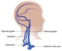
Vasculature Of The Head Texas Heart Institute from www.texasheart.org The first part extends from its origin to the medial border of the scalenus anterior muscle. Blood is received from the internal carotid artery and the basilar artery, which arises from the vertebral arteries. New blood vessel growth is called angiogenesis. Internal jugular vein • this is the larger of two vessels that drain blood from the head and neck into the subclavian. Major arteries veins and nerves of the body anatomy kenhub. The internal jugular moves alongside the internal carotid artery and receives blood from the brain. It extends on each side of the neck and divides at the level of the larynx into two branches: The posterior auricular, occipital and superficial temporal arteries (along with two branches of the internal carotid artery;
The subclavian artery is divided into three parts based on anatomical landmarks.
Blood vessels labeled head : There are three major types of blood vessels: Blood supply of the head and neck alila medical images. • capillaries, which enable the actual exchange of water and chemicals between the blood and the tissues. Blood is received from the internal carotid artery and the basilar artery, which arises from the vertebral arteries. Parotid gland, submandibular gland (right half), deep musculature (left half), lower jaw partially exposed, displaying blood vessels measures 9.4 x 7.1 x 9.4 weighs 2.65 lbs pack of 1 The head rests on the top part of the. These include the common carotid artery that carries blood from the heart to the brain. Internal jugular vein • this is the larger of two vessels that drain blood from the head and neck into the subclavian. The internal jugular moves alongside the internal carotid artery and receives blood from the brain. In the neck and head exterior to the skull, the external carotid artery provides blood flow to the skin, muscles, and organs. A web of blood vessels—arteries, veins, and capillaries—circulate blood to organs. The derivatives of the internal carotid arteries form the anterior blood supply (anterior circulation) of the brain, which includes the anterior and middle cerebral arteries.
Parotid gland, submandibular gland (right half), deep musculature (left half), lower jaw partially exposed, displaying blood vessels measures 94 x 71 x 94 weighs 265 lbs pack of 1 blood vessels labeled. Injuries to the scalp can cause excessive bleeding for various reasons: